What is the Learning Unit about?
In clinical practice, images not always speak for themselves. A chest X-ray, an endoscopic photograph, or a dermatoscopy image must be interpreted, and sometimes only a subtle deviation in an image is of importance. Besides the image interpretation itself, the information, weighed against a patient’s history, physical examination, and the clinician’s own reasoning. When it comes to image analysis, healthcare professionals need the critical skills to decide when and how much to trust an image analysis output. Learning Unit (LU) 103 introduces undergraduate students to AI-assisted image analysis. The LU combines a demonstration of what technology can do, but also includes active reasoning exercises. These focus on how to integrate AI output into clinical reasoning, and where might this tool be misleading?
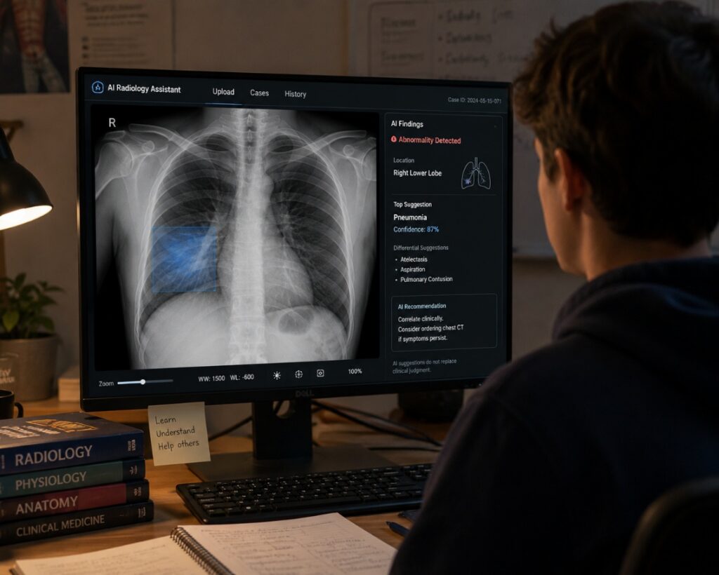
Students work through at least one Virtual Patient (VP) in which AI image analysis tools are embedded directly into the diagnostic workflow. One of them is Robert Baley, a 46-year-old firefighter who presents with four days of cough, fever, chills and sharp chest pain. After taking a history, performing a physical examination, and forming their own initial impression of the chest X-ray, students are then shown the output of an AI image analysis tool and must decide whether the AI finding changes their diagnosis.
Beyond the virtual patient cases, LU103 is supported by learning about the use of image analysis in four medical specialties, i.e. dermatology, radiology, cardiology and pathology. Besides presentations, working through and discussing clinical cases is part of the module. The clinical cases illustrate both failure modes but also success scenario, and includes structured multiple-choice questions suited to interactive polling tools. Failure cases cover topics such as population mismatch, dataset bias, and out-of-scope use, which are risks that are well-documented in the literature and directly relevant to students’ future practice.
How We Developed It
One of the first decisions we faced was how to handle AI tools in the learning design. AI in medical image analysis is already very mature compared to many other applications of AI in medicine. Validated tools exist across radiology, pathology, and dermatology, and some are already embedded in clinical workflows. Our intention was therefore to let students interact with real AI output, which would create a learning experience that they might encounter during their training and in their later career.
In practice, however, integrating live AI tools proved more complicated than anticipated. Access, licensing, and reproducibility all posed barriers. A tool that works in one institution’s system may not be available in another’s. Output can vary depending on image quality, acquisition parameters, and software version. This made it difficult to design a consistent learning experience around a tool students cannot reliably access themselves.
Our solution was to design AI output that is realistic rather than real. For the virtual patient cases, we created plausible AI analysis outputs that mirror the format and reasoning of actual clinical AI tools. This allowed us to control the pedagogical content precisely: to design AI output that sometimes agrees with the student’s own read, sometimes adds a new finding, and sometimes raises questions the student must consider. Rather than asking “what does the tool say?”, the learning task becomes “is this output trustworthy, and why?“
What Did We Learn?
Developing LU103 reinforced something that is exciting and challenging at the same time AI in medical image analysis is not a future technology. It is already here, but it is also still error prone. Learning about the tools, the application and the interpretation of findings is therefore crucial an very timely.
The cases we built that reflected potential errors in image analysis output (e.g. around population mismatch and dataset bias) are not hypothetical. These are documented problems with documented consequences. The tendency of models to perform well on the distribution they were trained on, but not as good when applied elsewhere, is one of the most important things students can learn about this technology. Framing it through a concrete clinical cases to practice with and discuss in a group, makes the stakes tangible in a way that abstract explanations cannot.
We also learned that the absence of a real tool does not have to mean an absence of authenticity. Carefully designed simulated AI output can be pedagogically rich, and logistically more feasible than access to a live tool. We were able to construct cases that reflect the tensions and ambiguities that are most important for learning. Students are not passive observers of what an algorithm produces, they are active reasoners who must account for what it might be missing.
Finally, this learning unit reflected a good balance between the benefits and the risks of the use of image analysis. Students often arrive with the assumption that AI in medicine is either dangerous or infallible. LU103 offers a third perspective: AI tools in image analysis can be genuinely useful, but they have well-characterized failure modes. Understanding those failure modes is itself a clinical competency.
Stay connected with D-CREDO and follow our journey on LinkedIn for more updates, insights, and stories.